Posterior Rib Cage Muscles / Internal Intercostals Learn Muscles : Posterior rib cage muscles :. Womens body parts stomach 4 photos of the womens body parts stomach body diagram stomach, body parts digestive system, body parts in stomach area, body parts liver, body parts spleen, human body parts stomach, woman body organs, woman body parts found, stomach, body diagram stomach, body parts digestive system, body. There are five muscles that make up the thoracic cage; The rectus abdominis runs between the ribs and the pubic bone and supports movements between the rib cage and the pelvis. Posterior rib cage muscles : Take five minutes out of your day to work on your posture and relieve achy muscles with these rib cage exercises.
The eleven pairs of internal intercostal muscles are found posterior to the external intercostals. The middle and upper part of your spine is called the thoracic region and it helps to support your upper body. Between each rib lie several layers of intercostal muscles that are responsible for expanding and shrinking. These muscles originate on the second through the 12 th ribs, and insert on ribs one to 11. Posterior rib cage muscles :
Internal Intercostal Muscles Attachments Supply Action Kenhub from thumbor.kenhub.com A rib has a flat body, as you can see from the picture of the anatomy of the human rib cage. The middle and upper part of your spine is called the thoracic region and it helps to support your upper body. Of all 24 ribs, the first seven pairs are often labeled as 'true.'. The transversus thoracic muscles originate from the posterior surface of the xiphoid process and the lower part of the body of the sternum. The human rib cage is made up of 12 paired rib bones; Muscles of the spine and rib cage | musculoskeletal key : These muscles may be located anteriorly, posteriorly, and/or laterally. Clench your glutes to hold your torso parallel to the floor.
Rib cage pain may be sharp, dull, or achy and felt at or below the chest or above the navel on either side.
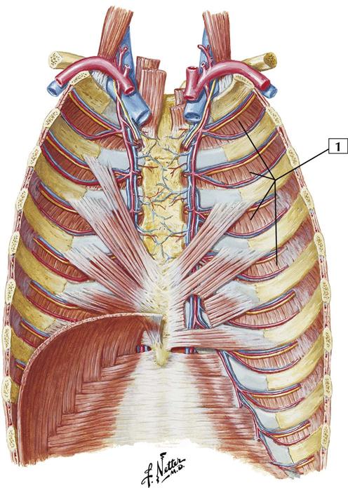
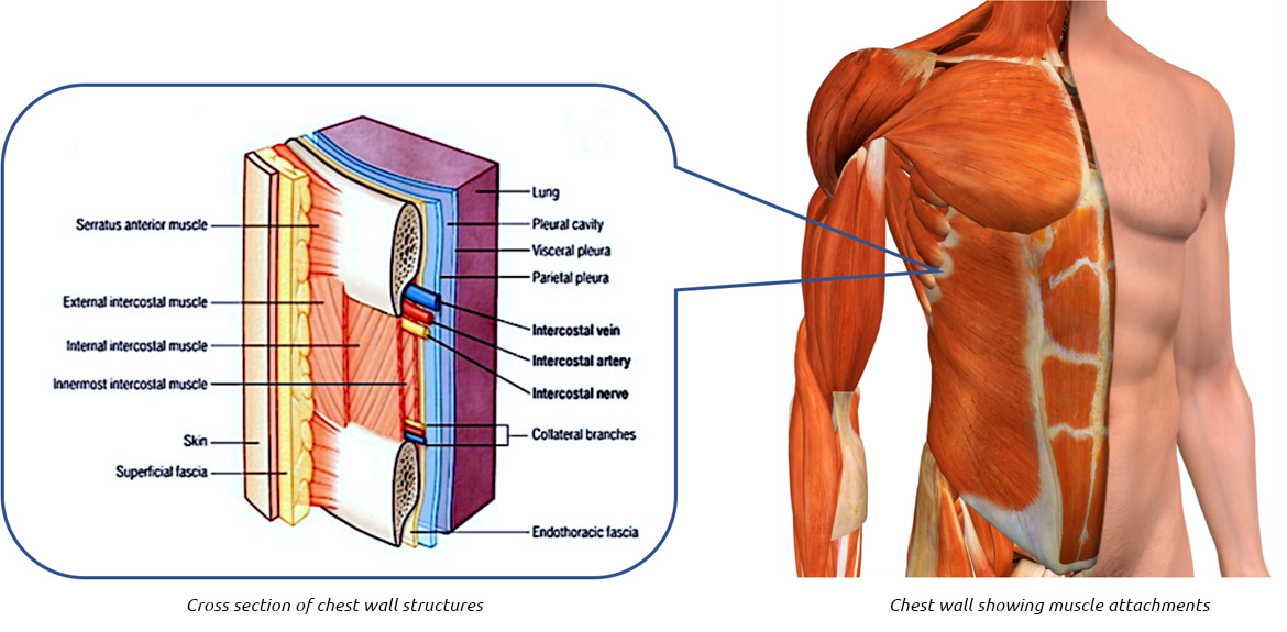
An inhalation is accomplished when the muscular diaphragm, at the floor of the thoracic cavity, contracts and flattens, while the contraction of intercostal muscles lift the rib cage up and out. There are five muscles that make up the thoracic cage; Between each rib lie several layers of intercostal muscles that are responsible for expanding and shrinking. Muscles of the rib cage muscles that move the rib cage attach to the rib cage. Rib cage pain can be caused. It is common for a client's back muscles to be excessively tight or crooked to compensate for rib pain. The other attachment of these muscles is usually considered to be either superior or inferior to the rib attachment. The eleven pairs of internal intercostal muscles are found posterior to the external intercostals. These muscles may be located anteriorly, posteriorly, and/or laterally. There are some other muscles that do not comprise the thoracic wall, but do attach to it. Grab a dumbbell and position yourself so you're perpendicular to the seat. The intercostals (external, internal and innermost), subcostals, and transversus thoracis. The intercostal muscles have different layers that are attached to the ribs to help build the chest wall and.
Lay back so your upper back is resting on the pad. The thoracic cage (rib cage) is the skeletal framework of the thoracic wall, which encloses the thoracic cavity. Posterior rib cage muscles : The fibres pass superolaterally to insert into the internal surface of costal cartilages of ribs two to six. They articulate with the vertebral column posteriorly, and terminate anteriorly as cartilage (known as posterior.
Thorax Cards 3 1 To 3 26 Basicmedical Key from basicmedicalkey.com This muscle assists in depression of the ribs. Injuries to your rib cage or muscles in your upper chest can cause rib pain ranging from a dull ache to sudden, sharp jabbing pains in the affected area. Issues with your muscles, ligaments, or ribs in your back can often cause rib pain in the back. The eleven pairs of internal intercostal muscles are found posterior to the external intercostals. Related posts of rib cage diagram with organs womens body parts stomach. The human rib cage is made up of 12 paired rib bones; Perform dumbbell pullovers to work the muscles along your rib cage. These muscles act to change the volume of the thoracic cavity during respiration.
Between each rib lie several layers of intercostal muscles that are responsible for expanding and shrinking.
The last two, the floating ribs, have their cartilages ending in the muscle in the abdominal wall. Each are symmetrically paired on a right and left side. Rib cage, therefore scm is considered an accessory muscle of respiration • medial to the scm lies the carotid sinus & carotid arteries; There are some other muscles that do not comprise the thoracic wall, but do attach to it. The intercostal muscles of the ribcage. Lay back so your upper back is resting on the pad. These muscles may be located anteriorly, posteriorly, and/or laterally. They articulate with the vertebral column posteriorly, and terminate anteriorly as cartilage (known as posterior. The fibres pass superolaterally to insert into the internal surface of costal cartilages of ribs two to six. The human rib cage is a component of the human respiratory system. Perform dumbbell pullovers to work the muscles along your rib cage. These muscles act to change the volume of the thoracic cavity during respiration. Grab a dumbbell and position yourself so you're perpendicular to the seat.
In those cases, deep tissue therapy to release tension in the back may help release the posterior ribs or compensation patterns. Womens body parts stomach 4 photos of the womens body parts stomach body diagram stomach, body parts digestive system, body parts in stomach area, body parts liver, body parts spleen, human body parts stomach, woman body organs, woman body parts found, stomach, body diagram stomach, body parts digestive system, body. The configuration of the lower five ribs gives freedom for the expansion of the lower part of the rib cage and for the movements of the diaphragm, which has an extensive origin from the rib cage and the vertebral column. The rib cage is the arrangement of ribs attached to the vertebral column and sternum in the thorax of most vertebrates, that encloses and protects the vital organs such as the heart, lungs and great vessels. Related posts of muscle anatomy rib cage muscle anatomy drawing.
Chest Wall Lumps Rib Injury Clinic from www.ribinjuryclinic.com The rectus abdominis runs between the ribs and the pubic bone and supports movements between the rib cage and the pelvis. Muscles of the rib cage muscles that move the rib cage attach to the rib cage. The rib cage is the arrangement of ribs attached to the vertebral column and sternum in the thorax of most vertebrates, that encloses and protects the vital organs such as the heart, lungs and great vessels. Muscle spasms felt within the rib cage may also be caused by the abdominal muscles. The fibres pass superolaterally to insert into the internal surface of costal cartilages of ribs two to six. On the interior wall of the rib body is a channel, sulcus costae, with blood vessels and nerves. These muscles act to change the volume of the thoracic cavity during respiration. The other attachment of these muscles is usually considered to be either superior or inferior to the rib attachment.
This muscle assists in depression of the ribs.
The intercostal muscles have different layers that are attached to the ribs to help build the chest wall and. Tuesday 2nd april bircher muesli , bee flies and a bit more about breathing. On the interior wall of the rib body is a channel, sulcus costae, with blood vessels and nerves. If all these muscles are tight, it can leave you feeling constricted. The thoracic cage (rib cage) is the skeletal framework of the thoracic wall, which encloses the thoracic cavity. Each are symmetrically paired on a right and left side. The major abdominal muscles include the transverse abdominals, the rectus abdominis, and the external and internal oblique muscles. Lay back so your upper back is resting on the pad. Related posts of muscle anatomy rib cage muscle anatomy drawing. The configuration of the lower five ribs gives freedom for the expansion of the lower part of the rib cage and for the movements of the diaphragm, which has an extensive origin from the rib cage and the vertebral column. Human muscles · april 17, 2020. These muscles originate on the second through the 12 th ribs, and insert on ribs one to 11. Anteriorly, most are attached directly to the sternum.
There are some other muscles that do not comprise the thoracic wall, but do attach to it rib cage muscles. Posterior rib cage muscles :
0 Comments:
Post a Comment